NC State’s New Standing CT a Game-Changer for Equine Care
Now, our equine experts can scan the heads and throat latches of horses without having to put the animals under anesthesia.
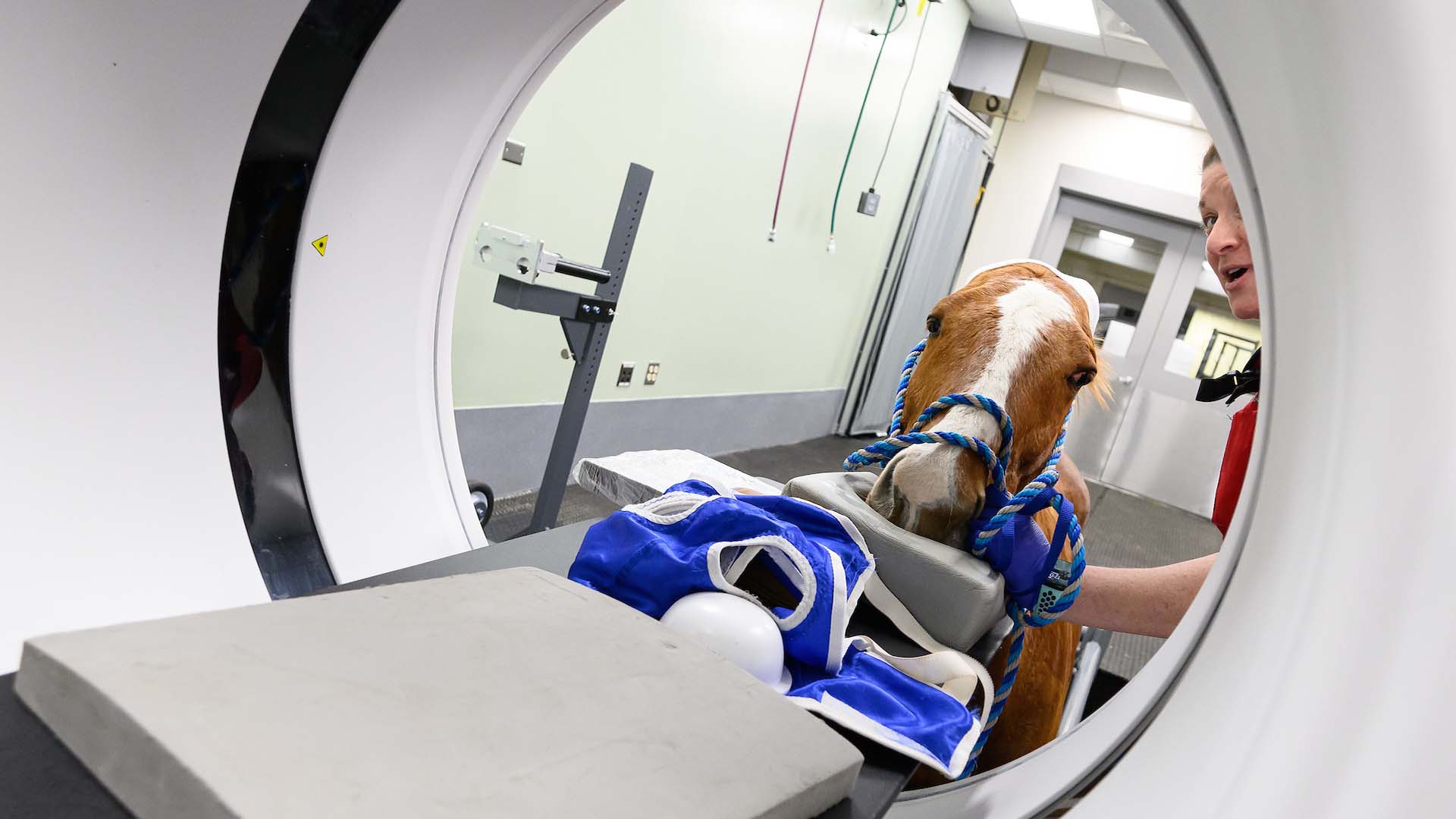
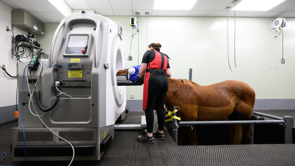
In its continuous quest to offer the best of veterinary care, the NC State Veterinary Hospital has installed standing CT equipment that allows our equine experts to scan the heads and throat latches of horses without having to put the animals under general anesthesia.
“Horses are big animals, and recovery from anesthesia is a big event with a lot of risk,” says Dr. Callie Fogle, professor of equine surgery. “One of the tough parts is that imaging, at least advanced imaging including CT and MRI, has been mostly done under general anesthesia, so this is a big step for us because we can now do imaging of the head and the front part of the horse standing, which then allows us to complete a surgical procedure standing as well. So we’re very excited to have this technology.”
The standing CT allows sedated horses to walk into stocks on a platform that then lowers the animal below the floor until its head is level with the scanner. The CT machine then moves back and forth over the horse’s head and neck.
To see how it works, click here.
Fogle estimates that there are fewer than 12 standing CT machines being used for veterinary purposes in the United States. NC State began offering the standing CT about a year ago.
In addition to keeping the animal from having to be anesthetized, the standing CT allows for three-dimensional imaging whereas radiographs supply two-dimensional images of 3D anatomical structures. The new machine helps doctors pinpoint specific lesions and problems so that surgeons can minimize risk to surrounding tissue.
“It will allow us to understand a number of problems that we also see following surgeries on the head much better,” says Dr. Timo Prange, associate professor of equine surgery. “So it will help us with the surgical planning … and then down the road you can also identify the cause for certain complications that we might now understand at this point.”
Prange also explained how the machine will improve learning for NC State College of Veterinary Medicine students, interns and residents.
“It’s fantastic,” he says. “The anatomy of the head is quite complicated. Now that we have the CT it is much easier for them to understand what we are going to do, what the problems are, how we can solve them. It’s an amazing tool, and since we can create three-dimensional images, it’s also a nice way of showing the owner of the animal what’s wrong and what we’re going to do.”
Dr. Steven Marks, director of the NC State Veterinary Hospital, says acquiring the standing CT equipment, made possible mostly through donations to the hospital, allows the college to expand its three-point mission to provide excellent patient care, to best educate the next generation of healers and to expand the limits of medicine based on scholarly activity.
In addition, state-of-the-art equipment helps NC State be a better partner to the community, which is also a priority for the college.
“I think it’s really important to recognize that the NC State Veterinary Hospital partners with veterinarians in the community, and we can provide services that are not available everywhere,” Marks says. “So it’s a partnership, it’s a team together, not just inside the veterinary hospital but with the outside clinicians, and we work together to provide the best patient care possible.”