Research Roundup March 2025
Not just filling in gaps, our researchers are getting to the source of both critical diseases and educational needs. Discover the latest exciting breakthroughs and impactful advancements made by our internationally recognized scientists at the NC State College of Veterinary Medicine.
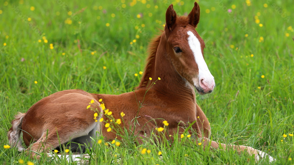
Evaluating the Significance of Vitamin D Levels in Critically Ill Foals
Kamr AM, Bartish C, Summers J, Horton J, Hostnik LD, Orr K, Browne N, Dembek KA, Saliba C, Gomez DE, Toribio RE.
Vitamin D is responsible for helping the intestines absorb a multitude of other vitamins and minerals like calcium, phosphorus and magnesium. It also promotes proper immune function and the integrity of the epithelial tissue that serves as a protective layer lining internal organs and body cavities. However, little is known about the signaling pathways of vitamin D and its role in the immune system of foals fighting infections, specifically critical cases like sepsis. In order to gain a clearer understanding, an international team of researchers, including faculty from the Department of Clinical Sciences, collaborated on a study that looked at the relationship between vitamin D levels and immune responses in newborn foals that were hospitalized, particularly focusing on antimicrobial peptides (which help fight infections) and other immune markers. The results indicated that lower levels of vitamin D and antimicrobial peptides are associated with more severe illness and higher mortality rates in foals, especially those with sepsis. The study suggests that vitamin D plays an important role in the immune system of foals and could impact their ability to fight infections, leading to improved treatments and management strategies.
The study was published in the Journal of Veterinary Internal Medicine and can be found here.
Finding the Links between Kidney Disease and Degenerative Joint Disease in Cats
Lascelles BDX, Ponnala R, Kamerling SG, Williams T.
Degenerative joint disease is a common condition in cats. Interestingly, almost 70% of cats with this condition also suffer from chronic kidney disease. Both of these conditions are known to be very painful and have limited treatment options available, making it a troublesome combination for pets, their concerned owners and treating veterinarians. In an effort to find new disease biomarkers and treatment options, a recent study led by Dr. Duncan Lascelles in the Department of Clinical Sciences analyzed the blood of 200 cats with varying levels of degenerative joint disease, pain and chronic kidney disease to better understand the underlying causes. The researchers found key biological pathways involved in degenerative joint disease and pain, such as inflammation and immune system activity. When chronic kidney disease was also present, additional pathways related to immune signaling were also identified. The findings suggest potential biomarkers for these diseases and could lead to new therapeutic targets. Additionally, because degenerative joint disease is also a major health issue in humans, these results may be relevant for human health studies.
The study was published in Frontiers in Pain Research and can be found here.
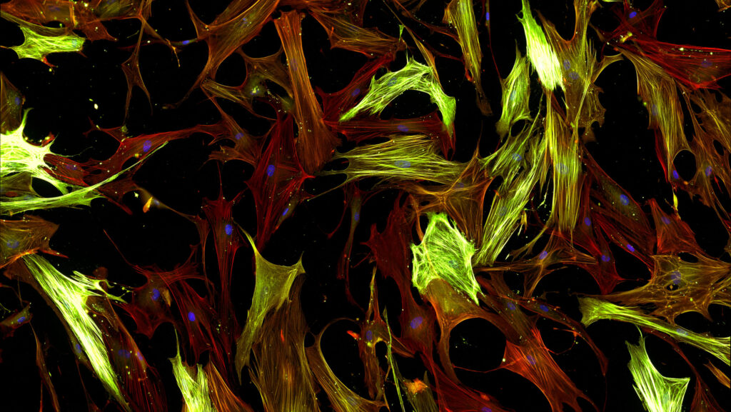
Studying Lung Cell Stiffness to Aid in Disease Research
Monaghan-Benson E, Aureille J, Guilluy C.
Tissue growth and homeostasis are processes our bodies depend on to stay healthy, and these processes rely on a number of factors within the cell microenvironment. However, it is still not well understood how our cells convert the mechanical signals from their surroundings into the biochemical signals that control their behavior. In an effort to discover more, a recent study that included Dr. Christophe Guilluy from the Department of Molecular Biomedical Sciences evaluated how the stiffness of the surrounding environment affected the survival of lung fibroblast cells, which are cells that help to produce and maintain the lung’s structural framework. Stiffness plays a crucial role in how cells sense and respond to their environment. The results found that when the cell environment is stiffer, the cells survive better due to increased activity of a protein called Ras. The activity triggers two important cell pathways that help the cells survive by affecting another protein, FOXO3a. When FOXO3a is turned off by Ras signaling, it reduces the levels of a protein called Bim, which promotes cell death. This entire signaling process helps cells stay alive on stiff surfaces and could help develop treatments for diseases like cancer and fibrosis, where stiffness in tissues plays a role.
The study was published in the Journal of Biological Chemistry and can be found here.
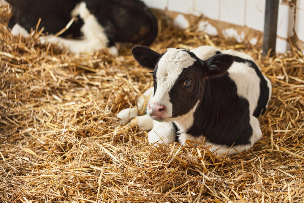
Addressing Intestinal Healing in Babies with Serious Infections
Rose EC, Simon JM, Gomez-Martinez I, Magness ST, Odle J, Blikslager AT, Ziegler AL.
Damage to the intestines and the barriers that protect them can lead to serious infections, like sepsis, without quick treatment and restoration of the epithelial barrier. Neonatal animals are more likely than older animals to have trouble healing this kind of damage, although it is unclear why. To answer this question, a team of multi-university researchers, including faculty from all three NC State College of Veterinary Medicine departments, conducted a study using a neonatal pig model to understand why neonates have a harder time than adults recovering from intestinal injury and to find potential treatments that could help improve recovery. The study found that the protein CSF-1 helps these intestinal cells heal more effectively but that it was active only in older pigs after an injury, not in babies. When the researchers added CSF-1 to the injured intestines of the neonatal pig model, healing improved. These findings suggest that CSF-1 could be a potential treatment to help babies recover better from intestinal injury.
The study was published in the American Journal of Physiology and can be found here.
Developing and Evaluating Animal Welfare Case Studies for Veterinary Students
Améndola L, Pairis-Garcia M, Millman S, Proudfoot KL.
Educational research is critical to the growth and improvement of the veterinary profession. Case studies are an important tool in veterinary education that provide students with a chance to strengthen their critical thinking and problem-solving skills in applicable scenarios. These skills are especially important surrounding complex and multidisciplinary topics like animal welfare that require a multitude of perspectives, sensitivity and adaptability. Although there is an abundance of literature and research on creating case studies for other educational purposes, there is not any specific guidance for developing effective case studies when it comes to animal welfare. A recent collaboration among faculty from multiple veterinary colleges, including Dr. Monique Pairis-Garcia from the Department of Population Health and Pathobiology, sought to establish a resource for veterinary educators to use when creating their own case studies, specifically studies on farm animal welfare. With the help of both curricular and content experts, the team created four case studies, with two versions of each case for a total of eight cases and formally outlined the process for developing and evaluating case studies that focus on farm animal welfare.
The article was published in the Journal of Veterinary Medical Education and can be found here.
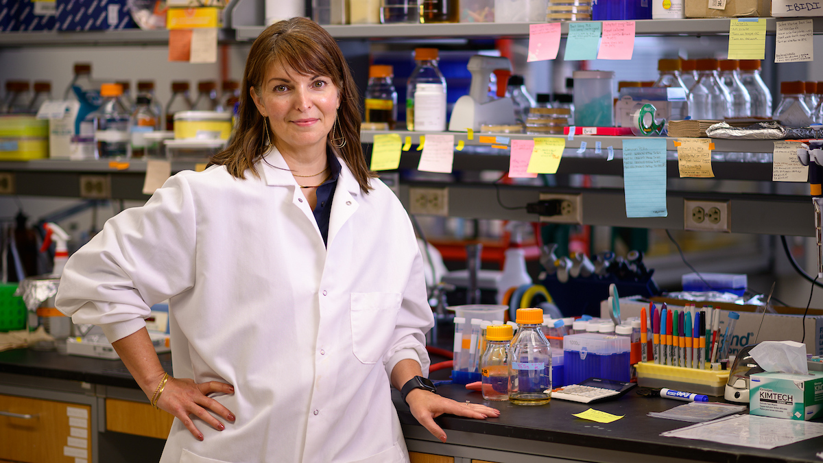
Research Connection: Dr. Kursten Pierce
It’s easy to forget that certain kinds of technology, such as X-ray imaging, put off potentially harmful radiation. To keep patients safe, lead gowns are often used to protect internal organs. But have you ever wondered who’s making sure that the doctors, staff and house officers using radiation technology are also protected?
Enter Dr. Kursten Pierce, one of two veterinary professionals in the world who has extensive, designated training in safety protocols for when radiation-producing techniques are used during Interventional Cardiology. In this practice, a technique called fluoroscopy is used, which is a real-time X-ray that works to image a patient’s heart. This ensures that life-saving heart procedures, such as pacemaker implantation and heart defect correction, can be performed.
Fluoroscopy produces significantly higher doses of radiation than a typical X-ray, so patients, doctors, technicians and staff need a higher level of protection. What does that look like? Watch to find out.
- Categories: