A Sharp Vision for Veterinary Ophthalmology

An advanced ocular imaging system coming to the NC State Veterinary Hospital strengthens the ophthalmology service’s unmatched treatment capabilities and commitment to personalized care.
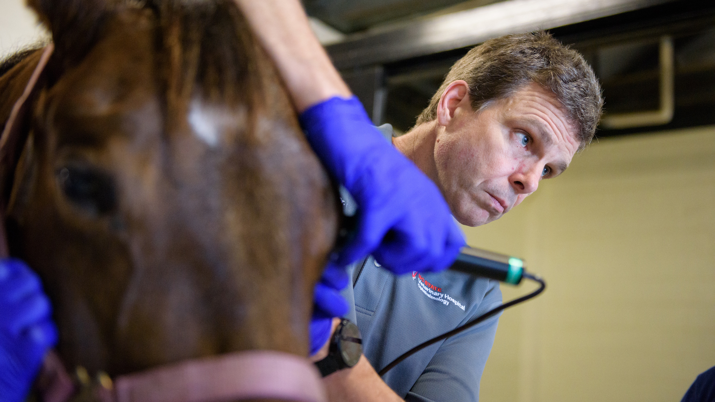
The new addition, a 70-megahertz ultrasound, specializes in detailed microscopic scans of the eye and uses sound waves to get real-time evaluations of anatomical structures. It is particularly effective when evaluating the cornea, which has the thickness of the head of a pin and allows light to pass through the eye so the retina can send an image to the brain.
The new imaging system, made possible by a generous gift from Fred and April Eshelman, joins a collection of advanced tools used by the ophthalmology service: a 50-megahertz ultrasound and optical coherence tomography. Both tools are non-invasive; the imaging is completed with little to no touching of the eye.
Scans are often used to determine whether there are abnormalities contributing to a disease that cannot be seen during a routine examination. This provides an ophthalmologist with a better guide on the likelihood of needing surgery versus medication. For example, a scan using these high-frequency systems on dogs with glaucoma can evaluate the part of the eye where fluid should drain from.
In horses with fungal corneal infections, “surgery to remove the infected cornea and graft is possible, but knowing how extensive the infection is can make an important difference when giving owners medical and surgical options,” said Brian Gilger, professor of ophthalmology.
The cutting-edge optical coherence tomography tool is similar to an ultrasound but uses light waves. It can map out the cornea or the retina, giving clinicians a layer-by-layer view of the structure.

Gilger said the machine gives a clinician more microscopic-level information and does not require contact of a probe to the eye. In cases involving a corneal ulcer, which is an abrasion or erosion of the layers of the cornea, the cornea of dog, cat or horse patients is very fragile. In these cases, the mere act of blinking or holding an eye open can lead to a traumatic rupture. When using the OCT, that pressure can be avoided since it doesn’t touch the eye. Gilger said the service hopes to purchase a new OCT instrument that provides sharper details and faster scans, further improving imaging capabilities for patients, especially those with retinal disease.
“When your patients are often scared and cannot speak for themselves, anything that can provide a large amount of information without great duress is a huge asset,” said Gilger.
Ocular imaging, said Gilger, is at the heart of treating eye diseases, and the ophthalmology service is applying these advanced systems, often used in human medicine, seamlessly to the world of veterinary ophthalmology. They are integral parts of the ophthalmology team’s place at the forefront of innovative approaches to ophthalmic medicine, surgery and research, he said.

As medicine evolves, Gilger said, each patient needs more specific evaluations and therapy. One-size-fits-all treatment programs may not be the answer to combating severe ocular diseases.
“We as humans have a special relationship with the animals in our care,” said Gilger. “They are our family members and our co-athletes. They can even be our own eyes when trained as guiding companions.
“The NC State ophthalmology team is pushing the art of ophthalmology to the next level by providing world-class medical care and advanced imaging for the benefit of these special animals and the people who care for them.”
For more information on the Gilger lab’s visionary ophthalmology, go here.
In order to continue providing the advanced level of care the NC State Veterinary Hospital offers each and every day, it is important to keep pace with changing and improving technology. Your support of the ophthalmology service will help to fund upgrades to ocular imaging systems that save lives and lead to brighter tomorrows.
~ Jordan Bartel/NC State Veterinary Medicine
- Categories:


