Yona the Bear Undergoes CT-scan at VTH
When administrators from the Museum of Life and Science in Durham, NC noticed their black bear, Yona, walking with a limp, they wasted no time in getting her examined by the Museum’s consulting veterinarians. The checkup indicated that Yona, who was found as a four-pound cub by Appalachian Bear Rescue, had an angular deformity in her right limb. A CT-scan was needed to help diagnose the specific issue.
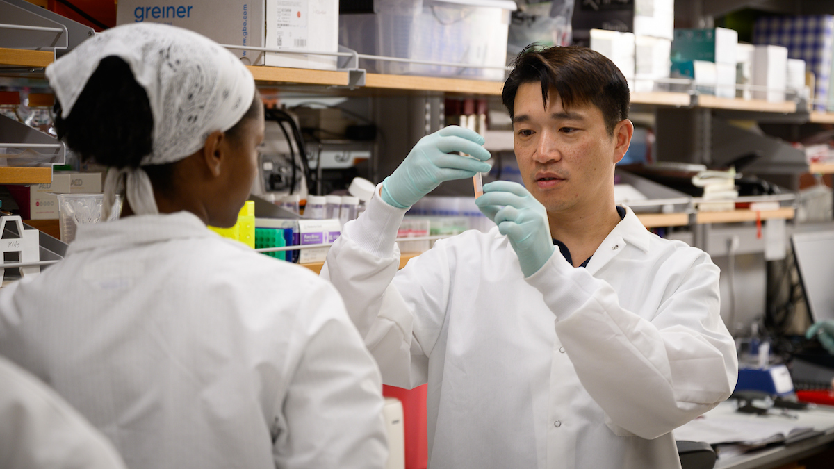
Arrangements were made with the Veterinary Teaching Hospital at North Carolina State University’s College of Veterinary Medicine and a team of clinicians, veterinary technicians, and museum staff assisted with the procedure. Led by Dr. Denis Marcellin-Little, a professor of orthopedics, the team included Dr. Michael Stoskopf, a professor of zoological medicine, and Dr. Kristen Messenger, who administered the anesthesia.
Yona—the name means “bear” in Cherokee— was sedated for about 2.5 hours and had a CT scan, radiographs, blood work, a dermatology exam, including skin scrape and punch biopsy, physical exam, extensive manipulation of her limbs, and more.
"It seems Yona injured her arm a long time ago,” Dr. Marcellin-Little said after studying all of the test results. “Fortunately, her arm is still growing. Her elbow is a little bit out of alignment, but that does not appear to be getting worse. She broke a piece of bone the size of an almond inside her elbow and that piece is moving around the joint. We’ll watch how she does and may remove the bone in the future as she grows, gains weight, and places more stress on the limb.”
Yona currently is doing just fine back in the bear habitat at the Museum of Life and Sciences. She continues to wrestle with Gus, the four year old black bear, swim in the bear pool, climb the mulberry tree, and relax near the waterfall—all to the delight of habitat visitors who come to see the bear that was in the news.
Watch Yona undergo a CT-scan in the NC State University report or the video on YouTube.
Posted June 24, 2010

Dr. Kristen Messenger administers additional sedatives before Yona undergoes a CT-scan at the College of Veterinary Medicine’s Veterinary Teaching Hospital at North Carolina State University. Photo by Wendy Savage.

Dr. Denis Marcellin-Little checks Yona’s positioning in the CT-scan. Dr. Marcellin-Little is a professor of orthopedics who asked by the Museum of Life and Scicence to consult on Yona’s condition. Photo by Wendy Savage.

Dr. Michael Stoskopf, a professor of zoological medicine, supervised Yona’s visit from arrival, sedation, and movement through the VTH including CT-scan, radiographs, blood work, and other activities. Photo by Wendy Savage.

Yona’s CT-scan shows a bone fragment the size of an almond in her right elbow joint. This has caused her elbow to be out of alignment and has resulted a limp. Museum officials will watch Yona as she grows to see if and when surgery will be appropriate to remove the bone fragment.