NIH Funds CVM Research into Genetic Causes of Intestinal Deformity
Dr. Nanette Nascone-Yoder, a research scientist with the NC State University College of Veterinary Medicine (CVM), has received a five-year, $1.5 million grant from the National Institutes of Health to study the genes responsible for intestinal formation. This work may lead to improved diagnosis and prevention of intestinal malrotation—or twisting—in human infants.
An assistant professor of developmental biology in the CVM Department of Molecular Biomedical Sciences, Dr. Nascone-Yoder and her research team have previously found the method by which the “gut tube”—the structure in vertebrate embryos that becomes the gastrointestinal tract—changes from a short, solid cylinder into an elongated hollow structure that loops and coils. The research paves the way toward greater understanding of how the initial asymmetric, or uneven, lengthening of the gut tube in an embryo drives the process of rotation and looping that must occur for intestines to form correctly.
“We know that the gut tube lengthens asymmetrically, and that these asymmetries are purposeful—defined in such a way as to lead to exact looping and rotation,” Dr. Nascone-Yoder says. “What we don’t know is the exact mechanism involved—how different genes and molecules interact in order to cause these specific asymmetries.”
Dr. Nascone-Yoder’s next steps will be to look at the genes and molecules responsible for controlling the process of elongation, particularly at a transcription factor known as Pitx2, which is required for normal asymmetric elongation of the gut tube.
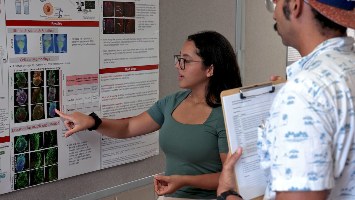
The $1.5 million NIH study builds upon earlier research funded by the National Science Foundation and published in Developmental Dynamics. In this research, the CVM team utilized frog embryos to study the earliest stages of gastrointestinal development. The frog’s transparent embryos allow researchers to observe structural changes in real time. For example, the frog gut tube increases its length by a factor of three in one 24-hour period, and the elongated structure also narrows and coils during that time.
The team wanted to know how the gut tube got longer, since there was no evidence that cell proliferation, or division, was responsible. They discovered that Rho GTPase, a molecule important to gastrulation—the process whereby an embryo rearranges cells to form layers that will eventually become the different tissues of the body—was telling the gut tube to get longer and thinner.
Under the influence of Rho GTPase and associated molecules, the cells inside the gut tube change shape and position. Then the interior cells push between the cells on the outside, forcing the tube to become both long and hollow.
“We’ve known that this molecule is important to cell migration and adhesivity,” says Dr. Nascone-Yoder. “It’s important to the earliest stages of embryonic development since it tells cells where and how to line up, so that other developmental processes can begin. Now we see that the same thing is happening here, with an organ.
“Approximately one in 500 human infants is born with intestinal malrotation—a common condition, which poses significant risk for life-threatening complications,” Dr. Nascone-Yoder continues. “Doctors are good at fixing problems after the fact, but no one has paid much attention to how the gastrointestinal tract forms to begin with. If we understand how the genes operate that are responsible for a gastrointestinal tract to form properly, it will give us insight into the causes of a host of common digestive organ birth defects.”
October 14, 2010