Alice the Chicken: Diagnosed with Uterine Cancer

Article by Whitney L.J. Howell
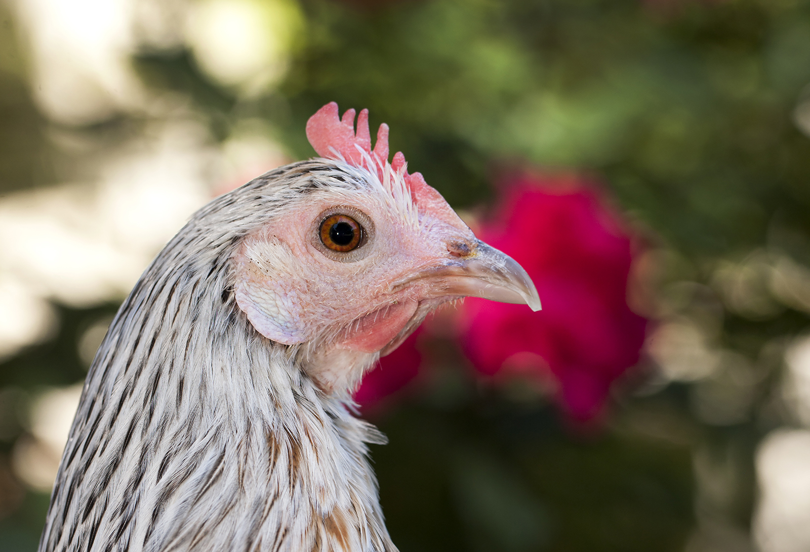
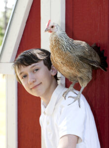
It’s said there’s often one hen to rule the roost. In the case of Alec Bergin, a 13-year-old boy from Moore County, that hen is Alice, a rare breed Phoenix chicken. Ever since Alice joined the Bergin family with three other Phoenix hens, Alec has hand-fed her treats and watched her assume a leading-lady role, hatching and mothering her share of 12 chicks. “This is Alec’s own flock, and he takes care of them,” says Jennifer Bergin, Alec’s mother. “He’s responsible for feeding and watering them. He goes outside and spends 20 minutes every day just watching them to make sure they’re acting normally. If anything’s wrong, he can catch it early on.” And that’s exactly what Alec did one evening. Instead of running for her treat like normal, Alice stayed on her nest. She only half-heartedly pecked at the niblet, and after looking her over, Alec and his mother noticed her distended belly and discovered her back end was covered in feces. Their first assumption: she couldn’t lay her eggs.

Taking Alice to the community veterinarian wasn’t an option—chickens aren’t everyday pets. To get this family hen the proper care, Bergin brought her to the NC State Veterinary Hospital and put her in Jeff Applegate’s hands.“When Alice came in, she was very lethargic and exhibited the distended belly or coelom so we started with a physical exam, completed blood work, and proceeded to complete an emergency ultrasound in concert with the Radiology Service,” says Dr. Applegate, a clinical veterinarian specializing in companion exotic animal medicine. “The ultrasound revealed significant fluid and abnormal tissue in and around the reproductive tract,” Applegate continues. “There shouldn’t have been any free fluid in her belly. Of the more routine birds that we treat as pets, the abdomen or more appropriately referred to as a coelom can be described as a central column of organs like the heart, liver, and intestines, with the remaining space occupied by the surrounding air sacs and lungs. ” Reproductive disease is common in chickens, and it’s analogous to uterine disease in humans and other mammals. The ultrasound showed Alice had free coelomic fluid and abnormal tissue in her oviduct, the tunnel in which an egg forms and by which it leaves the hen’s body. The diagnosis was oviductal adenocarcinoma—Alice had uterine cancer. The treatment: a salpingohysterectomy, the avian equivalent to spay.
[pullquote color=”orange”]“She’s a valued member of our family as much as the cats and dogs are.” [/pullquote]
Once the Bergins green-lighted surgery, understanding Alice would never again lay eggs, Applegate assembled a team from the Exotic Animal Medicine Service to combine their skills during Alice’s operation. Pooling talents from multiple specialties is a benefit the NC State Veterinary Hospital offers patients according to Applegate. In cases like this, many collaborating services may include the emergency and critical care service, radiology, anesthesia, and surgery. The surgery—an invasive procedure with the surgeons removing Alice’s diseased oviduct through a small L-shaped incision behind her left leg—was a success with few complications and a moderate amount of bleeding. After two weeks recuperating in the Bergin’s master bathroom, Alice moved back outside and assumed her leadership position. “She’s living with friends and doesn’t look any different from the other hens,” Bergin says. “She’s a valued member of our family as much as the cats and dogs are.”
[divider thickness=5%]
[section type=”full” id=”optional” color=”gray”] [feature_title color=”black”]Surgeon’s Notes[/feature_title]
[feature_image align=”left” title=”” src=”https://news.cvm.ncsu.edu/wp-content/uploads/sites/3/2015/09/oviduct-Alice.jpeg” linkto=”https://cvm.ncsu.edu/research/departments/docs/programs/exotic/” alt=”Ultrasound of Alice”]The decision to pursue an avian salpingohysterectomy is often considered a last resort due to risks associated with avian anesthesia and surgery. Veterinarians who specialize in avian medicine tend to be comfortable with these types of procedures but that by no means eliminates the risks. Effort taken prior to surgery ensure that the patient is as stable as possible for anesthesia. These efforts include conducting a thorough physical exam, blood work, and imaging such as radiographs (X-rays) and/or ultrasound to characterize the current disease process as thoroughly as possible and to rule out any other underlying diseases. In Alice’s case, she was also administered a medication by injection weeks prior to the surgery to help reduce any reproductive activity and decrease bleeding during surgery.
Avian patients that undergo surgery at NC State through the Exotic Animal Medicine Service are managed with the most advanced techniques possible. Each patient is intubated with a tube in their airway to manage breathing, an intravenous catheter is placed in a vein for fluid support, and they are monitored with a variety of equipment including an ECG to monitor heart rate and rhythm, a pulse oximeter to monitor the oxygen in the blood, a capnograph to monitor exhaled carbon dioxide, and equipment for temperature and blood pressure monitoring. Specialized surgical instrumentation are employed such as custom made forceps, radiosurgery and lenses worn by the surgeon to magnify the surgical field. Following surgery, avian patients are hospitalized in one of a variety hospital wards each specializing in a different level of care, from general hospital to the intensive care unit.
[/feature_image]
[/section]
- Categories:


